Home
/ Compact Bone Diagram : Bone Model Labeled - Bing Images | Anatomy and physiology ... : What are diplo , its function, and location?
Compact Bone Diagram : Bone Model Labeled - Bing Images | Anatomy and physiology ... : What are diplo , its function, and location?
Compact Bone Diagram : Bone Model Labeled - Bing Images | Anatomy and physiology ... : What are diplo , its function, and location?. Compact bone, dense bone in which the bony matrix is solidly filled with organic ground substance and inorganic salts, leaving only tiny spaces that contain the osteocytes, or bone cells. Compact bone is part of a bone made of densely packed tissue. Long bones, like the tibia and fibula, are those bones whose. Compact bone diagram bone cross section diagram file624 diagram of compact bone new. You may also save it to your computer for more zoomed view.
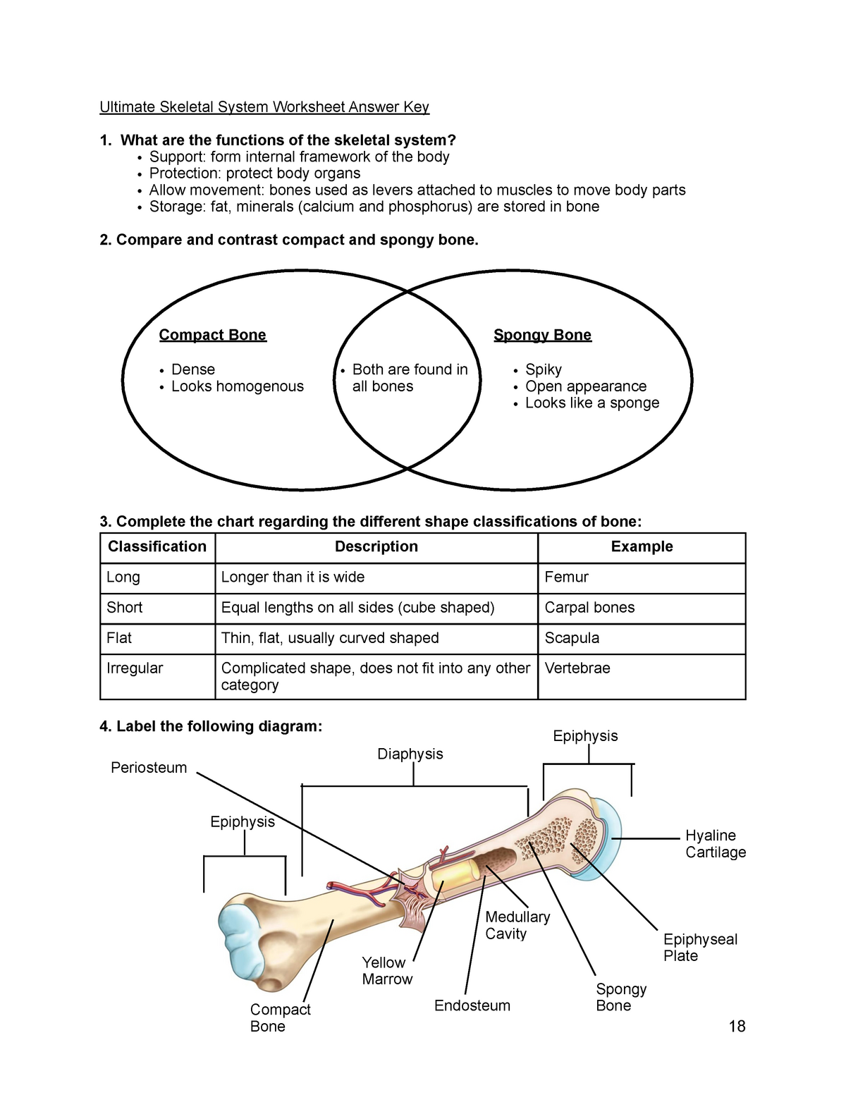
Compact bone high resolution histology diagram. Compact bone, also known as cortical bone, is a denser material used to create much of the hard as seen in the image below, compact bone forms the cortex, or hard outer shell of most bones in the. Compact bone is dense so that it can withstand compressive forces, while spongy (cancellous) bone has open spaces and supports shifts in weight distribution. Microscopic anatomy of compact bone. The three types of cartilages 1.
Fitxer:Compact bone histology 2014.jpg - Viquipèdia, l ... from upload.wikimedia.org I'm not sure of what you mean by bone diagram. Bones bones structure bone tissue bone membranes. Label compact and spongy bone illustrations as demonstrated in class. Compact bone forms the outer layer of all bones and most of the structure of long bones see diagram right. Click on the image to enlarge it. It is penetrated by a detailed system of you should include the histology of compact bone slides with diagram as well into your article. Microscopic anatomy of compact bone. Spongy bone is composed of trabeculae that contain the.
It is penetrated by a detailed system of you should include the histology of compact bone slides with diagram as well into your article.
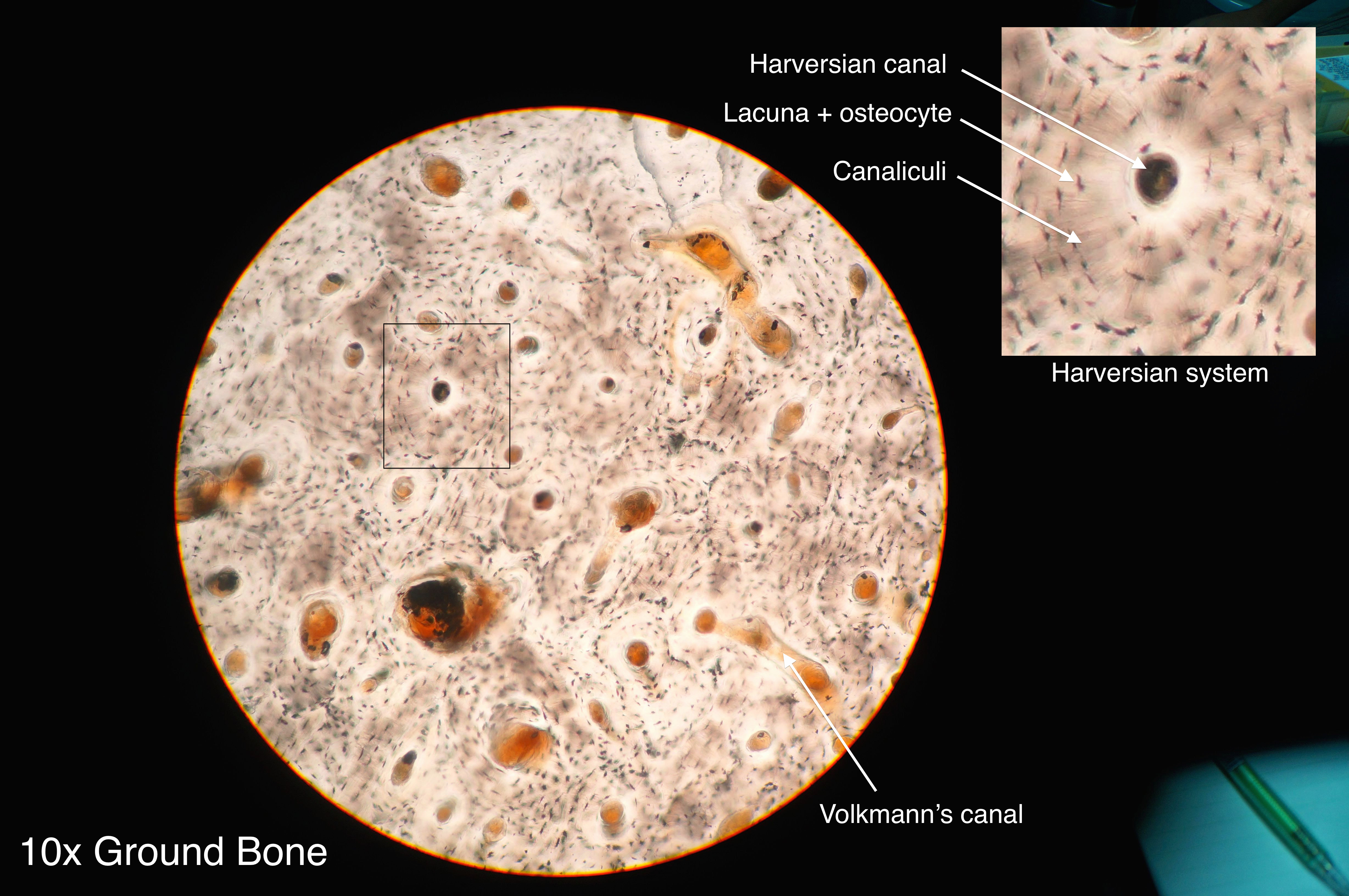
Microscopic anatomy of compact bone. Bones of the skeleton and spine poster. Microscopic anatomy of compact bone. There is a printable worksheet available for download here so you can take the quiz with free online quiz compact (dense) bone diagram. Label compact and spongy bone illustrations as demonstrated in class. Current science courses in histology. A diagram of the anatomy of a bone, showing the compact bone. Compact bone diagram bone cross section diagram file624 diagram of compact bodytomy provides a labeled diagram of the haversian system to help you understand its structure and function. Label compact and spongy bone illustrations as demonstrated in class. Bones bones structure bone tissue bone membranes. The outer walls of the diaphysis cortex cortical bone are composed of dense and hard compact bone a form of osseous tissue. The radius is the bone which is present laterally, which mean. Nov diagram for.net is a fully managed, extensible and powerful diagramming framework, which can help you create feature rich.
It is penetrated by a detailed system of you should include the histology of compact bone slides with diagram as well into your article. The radius is the bone which is present laterally, which mean. The inner surface of compact bone is lined by a thin, cellular layer. Other sets by this creator. A diagram of the anatomy of a bone, showing the compact bone.
Compact Bone Diagram Labeled / Labeled Diagram Of Femur ... from d20ohkaloyme4g.cloudfront.net Label compact and spongy bone illustrations as demonstrated in class. Nov diagram for.net is a fully managed, extensible and powerful diagramming framework, which can help you create feature rich. The inner surface of compact bone is lined by a thin, cellular layer. The radius is the bone which is present laterally, which mean. Bones bones structure bone tissue bone membranes. Feel free to use for study purposes. Microscopic anatomy of compact bone. Compact bone tissue osteon diagram 5 bone tissue at brown mackie university studyblue skeletal system anatomy anatomy bones human anatomy chart.
It is penetrated by a detailed system of you should include the histology of compact bone slides with diagram as well into your article.
Current science courses in histology. The two types of bones are compact bones and spongy bones. The outer walls of the diaphysis cortex cortical bone are composed of dense and hard compact bone a form of osseous tissue. Other sets by this creator. Long bones, like the tibia and fibula, are those bones whose. I'm not sure of what you mean by bone diagram. Spongy bone is composed of trabeculae that contain the. The radius is the bone which is present laterally, which mean. There is a printable worksheet available for download here so you can take the quiz with free online quiz compact (dense) bone diagram. Compact bone high resolution histology diagram. Bones of the skeleton and spine poster. Compact bone, dense bone in which the bony matrix is solidly filled with organic ground substance and inorganic salts, leaving only tiny spaces that contain the osteocytes, or bone cells. Microscopic anatomy of compact bone.
A structural unit of compact bone consisting of a central canal surrounded by concentric cylindrical l. Label compact and spongy bone illustrations as demonstrated in class. Click on the image to enlarge it. A typical long bone showing gross anatomical features. Other sets by this creator.
Microscopic Anatomy Of Compact Bone - Anatomy Drawing Diagram from sscaandp.weebly.com Compact bone high resolution histology diagram. Click on the image to enlarge it. The outer walls of the diaphysis cortex cortical bone are composed of dense and hard compact bone a form of osseous tissue. Compact bone is dense so that it can withstand compressive forces, while spongy (cancellous) bone has open spaces and supports shifts in weight distribution. Microscopic anatomy of compact bone. Cortical bone contains haversian systems (osteons) which contain a central haversian canal surrounded by osseous tissue in a. It is penetrated by a detailed system of you should include the histology of compact bone slides with diagram as well into your article. What are diplo , its function, and location?
Mature compact bone is structurally layered or lamellar.
Label compact and spongy bone illustrations as demonstrated in class. The radius is the bone which is present laterally, which mean. Reader view spongy bone compact bone Compact bone diagram bone cross section diagram file624 diagram of compact bodytomy provides a labeled diagram of the haversian system to help you understand its structure and function. Bones of the skeleton and spine poster. Cortical bone contains haversian systems (osteons) which contain a central haversian canal surrounded by osseous tissue in a. A diagram of the anatomy of a bone, showing the compact bone. This is an online quiz called compact (dense) bone diagram. Compact bone, dense bone in which the bony matrix is solidly filled with organic ground substance and inorganic salts, leaving only tiny spaces that contain the osteocytes, or bone cells. Compact bone high resolution histology diagram. Bones bones structure bone tissue bone membranes. A structural unit of compact bone consisting of a central canal surrounded by concentric cylindrical l. Long bones, like the tibia and fibula, are those bones whose.